- Giant-cell arteritis
-
Giant-cell arteritis Classification and external resources 
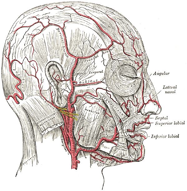
The arteries of the face and scalp.ICD-10 M31.5-M31.6 ICD-9 446.5 OMIM 187360 DiseasesDB 12938 eMedicine neuro/592 MeSH D013700 Giant-cell arteritis (GCA or Temporal arteritis or Cranial arteritis) or Horton disease is an inflammatory disease of blood vessels most commonly involving large and medium arteries of the head. It is a form of vasculitis.
The name (giant cell arteritis) reflects the type of inflammatory cell involved[1] as seen on a biopsy.
The terms "giant-cell arteritis" and "temporal arteritis" are sometimes used interchangeably, because of the frequent involvement of the temporal artery. However, it can involve other large vessels (such as the aorta in "giant-cell aortitis"[2]). Giant-cell arteritis of the temporal artery is referred to as "temporal arteritis," and is also known as "Cranial arteritis" and "Horton's disease."[3]:840
Contents
Signs and symptoms
It is more common in males than females by a ratio of 2:1. The mean age of onset is > 50 years, and it is rare in those less than 50 years of age.
Patients present with:
- bruits
- fever
- headache[4]
- tenderness and sensitivity on the scalp
- jaw claudication (pain in jaw when chewing)
- tongue claudication (pain in tongue when chewing) and necrosis[5][6]
- reduced visual acuity (blurred vision)
- acute visual loss (sudden blindness)
- diplopia (double vision)
- acute tinnitus (ringing in the ears)
The inflammation may affect blood supply to the eye and blurred vision or sudden blindness may occur. In 76% of cases involving the eye, the ophthalmic artery is involved causing arteritic anterior ischemic optic neuropathy.[7] Loss of vision in both eyes may occur very abruptly and this disease is therefore a medical emergency.
Associated conditions
The disorder may coexist (in one quarter of cases) with polymyalgia rheumatica (PMR), which is characterized by sudden onset of pain and stiffness in muscles (pelvis, shoulder) of the body and is seen in the elderly. GCA and PMR are so closely linked that they are often considered to be different manifestations of the same disease process. Other diseases related with temporal arteritis are systemic lupus erythematosus, rheumatoid arthritis and severe infections.
Diagnosis
Physical exam
- Palpation of the head reveals prominent temporal arteries with or without pulsation.
- The temporal area may be tender.
- Decreased pulses may be found throughout the body.
- Evidence of ischemia may be noted on fundal exam.
Laboratory tests
- LFTs, liver function tests, are abnormal particularly raised ALP- alkaline phosphatase
- Erythrocyte sedimentation rate, an inflammatory marker, >60 mm/hour (normal 10–40 mm/hour), but may be normal in approximately 20% of cases.
- C-reactive protein, another inflammatory marker, is also commonly elevated.
- Platelets may also be elevated.
Biopsy
The gold standard for diagnosing temporal arteritis is biopsy, which involves removing a small part of the vessel and examining it microscopically for giant cells infiltrating the tissue. Since the blood vessels are involved in a patchy pattern, there may be unaffected areas on the vessel and the biopsy might have been taken from these parts. Unilateral biopsy of a 1.5–3 cm length is 85-90% sensitive (1 cm is the minimum).[8] So, a negative result does not definitely rule out the diagnosis. Thus, currently biopsy is only considered confirmatory for the clinical diagnosis, or one of diagnostic criteria.[6]
Imaging studies
Radiological examination of the temporal artery with ultrasound yields a halo sign. Contrast enhanced brain MRI and CT is generally negative in this disorder. Recent studies have shown that 3T MRI using super high resolution imaging and contrast injection can non-invasively diagnose this disorder with high specificity and sensitivity.[9]
Treatment
Corticosteroids, typically high-dose prednisone (40–60 mg. once daily), must be started as soon as the diagnosis is suspected (even before the diagnosis is confirmed by biopsy) to prevent irreversible blindness secondary to ophthalmic artery occlusion. Steroids do not prevent the diagnosis from later being confirmed by biopsy, although certain changes in the histology may be observed towards the end of the first week of treatment and are more difficult to identify after a couple of months.[10] The dose of prednisone is lowered after 2–4 weeks, and slowly tapered over 9–12 months. Oral steroids are at least as effective as intravenous steroids,[11] except in the treatment of acute visual loss where intravenous steroids appear to offer significant benefit over oral steroids [12]
References
- ^ "giant cell arteritis" at Dorland's Medical Dictionary
- ^ Walter MA, Melzer RA, Graf M, Tyndall A, Müller-Brand J, Nitzsche EU (May 2005). "[18FFDG-PET of giant-cell aortitis"]. Rheumatology (Oxford) 44 (5): 690–1. doi:10.1093/rheumatology/keh551. PMID 15728420. http://rheumatology.oxfordjournals.org/cgi/pmidlookup?view=long&pmid=15728420.
- ^ James, William D.; Berger, Timothy G.; et al. (2006). Andrews' Diseases of the Skin: clinical Dermatology. Saunders Elsevier. ISBN 0-7216-2921-0.
- ^ Moutray TN, Williams MA, Best JL (August 2008). "Suspected giant cell arteritis: a study of referrals for temporal artery biopsy" (PDF). Can. J. Ophthalmol. 43 (4): 445–8. doi:10.1139/i08-070. PMID 18711459. http://pubs.nrc-cnrc.gc.ca/cjo/cjo43/i08-070.pdf.
- ^ Sainuddin S, Saeed NR (December 2008). "Acute bilateral tongue necrosis – a case report". Br J Oral Maxillofac Surg 46 (8): 671–2. doi:10.1016/j.bjoms.2008.03.027. PMID 18499311.
- ^ a b Zadik Y, Findler M, Maly A, et al. (January 2011). "A 78-year-old woman with bilateral tongue necrosis". Oral Surg Oral Med Oral Pathol Oral Radiol Endod 111 (1): 15–9. doi:10.1016/j.tripleo.2010.09.001. PMID 21176820. http://www.sciencedirect.com/science?_ob=ArticleURL&_udi=B6WP1-51RVMB0-F&_user=10&_coverDate=01%2F31%2F2011&_rdoc=15&_fmt=high&_orig=browse&_origin=browse&_zone=rslt_list_item&_srch=doc-info(%23toc%236977%232011%23998889998%232814774%23FLA%23display%23Volume)&_cdi=6977&_sort=d&_docanchor=&_ct=111&_acct=C000050221&_version=1&_urlVersion=0&_userid=10&md5=9a2b15759358123bfbffedf7a8652add&searchtype=a.
- ^ Hayreh (April 3, 2003). "Ocular Manifestations of GCA". University of Iowa Health Care. http://webeye.ophth.uiowa.edu/dept/GCA/04-ocular.htm. Retrieved 2007-10-15.
- ^ Ypsilantis E, et al. (November 2011). "Importance of specimen length during temporal artery biopsy". Br J Surg 98 (11): 1556–1560. doi:10.1002/bjs.7595/abstract.
- ^ Bley TA, Uhl M, Carew J, et al. (October 2007). "Diagnostic value of high-resolution MR imaging in giant cell arteritis". AJNR Am J Neuroradiol 28 (9): 1722–7. doi:10.3174/ajnr.A0638. PMID 17885247. http://www.ajnr.org/cgi/pmidlookup?view=long&pmid=17885247.
- ^ Font RL, Prabhakaran VC (2007). "Histological parameters helpful in recognising steroid-treated temporal arteritis: an analysis of 35 cases". The British journal of ophthalmology 91 (2): 204–9. doi:10.1136/bjo.2006.101725. PMID 16987903.
- ^ "BestBets: Steroids and Temporal Arteritis". http://www.bestbets.org/bets/bet.php?id=708.
- ^ Chan CC, Paine M, O'Day J (September 2001). "Steroid management in giant cell arteritis". Br J Ophthalmol 85 (9): 1061–4. doi:10.1136/bjo.85.9.1061. PMC 1724128. PMID 11520757. http://bjo.bmj.com/cgi/pmidlookup?view=long&pmid=11520757.
External links
- Giant Cell Arteritis article at University of Iowa
- Polymyalgia rheumatica article from National Institute of Arthritis and Musculoskeletal and Skin Diseases
- Temporal (Giant Cell) Arteritis - PrimeHealthChannel.com
- PMRGCAuk - UK charity offering support, raising awareness, promoting research
Vasculitis/arteritis: systemic vasculitis (M30–M31, 446) Large vessel Takayasu's arteritis · Giant-cell arteritisMedium vessel Small vessel Pauci-immuneHypersensitivity vasculitis/Henoch–Schönlein purpuraUngroupedAcute hemorrhagic edema of infancy · Bullous small vessel vasculitis · Cutaneous small-vessel vasculitisOther Goodpasture's syndrome · Sneddon's syndromeCategories:- Rheumatology
- Diseases of the eye and adnexa
- Neurological disorders
- Medical emergencies
- Vascular-related cutaneous conditions
Wikimedia Foundation. 2010.