- Fluorescence in situ hybridization
-
 A metaphase cell positive for the bcr/abl rearrangement (associated with chronic myelogenous leukemia) using FISH. The chromosomes can be seen in blue. The chromosome that is labeled with green and red spots (upper left) is the one where the wrong rearrangement is present.
FISH (fluorescence in situ hybridization) is a cytogenetic technique developed by Christoph Lengauer that is used to detect and localize the presence or absence of specific DNA sequences on chromosomes. FISH uses fluorescent probes that bind to only those parts of the chromosome with which they show a high degree of sequence complementarity. Fluorescence microscopy can be used to find out where the fluorescent probe bound to the chromosomes. FISH is often used for finding specific features in DNA for use in genetic counselling, medicine, and species identification. FISH can also be used to detect and localize specific mRNAs within tissue samples. In this context, it can help define the spatial-temporal patterns of gene expression within cells and tissues.
Contents
Probes
 Urothelial cells marked with four different probes.
Probes are often derived from fragments of DNA that were isolated, purified, and amplified for use in the Human Genome Project. The size of the human genome is so large, compared to the length that could be sequenced directly, that it was necessary to divide the genome into fragments. (In the eventual analysis, these fragments were put into order by digesting a copy of each fragment into still smaller fragments using sequence-specific endonucleases, measuring the size of each small fragment using size-exclusion chromatography, and using that information to determine where the large fragments overlapped one another.) To preserve the fragments with their individual DNA sequences, the fragments were added into a system of continually replicating bacteria populations. Clonal populations of bacteria, each population maintaining a single artificial chromosome, are stored in various laboratories around the world. The artificial chromosomes (BAC) can be grown, extracted, and labeled, in any lab. These fragments are on the order of 100 thousand base-pairs, and are the basis for most FISH probes.
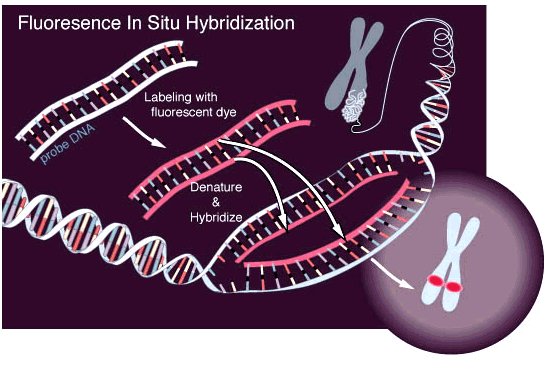
Preparation and Hybridization Process
 Scheme of the principle of the FISH Experiment to localize a gene in the nucleus.
First, a probe is constructed. The probe must be large enough to hybridize specifically with its target but not so large as to impede the hybridization process. The probe is tagged directly with fluorophores, with targets for antibodies or with biotin. Tagging can be done in various ways, such as nick translation, or PCR using tagged nucleotides.
Then, an interphase or metaphase chromosome preparation is produced. The chromosomes are firmly attached to a substrate, usually glass. Repetitive DNA sequences must be blocked by adding short fragments of DNA to the sample. The probe is then applied to the chromosome DNA and incubated for approximately 12 hours while hybridizing. Several wash steps remove all unhybridized or partially-hybridized probes. The results are then visualized and quantified using a microscope that is capable of exciting the dye and recording images.
If the fluorescent signal is weak, amplification of the signal may be necessary in order to exceed the detection threshold of the microscope. Fluorescent signal strength depends on many factors such as probe labeling efficiency, the type of probe, and the type of dye. The dye incorporation rate can be quantified photometric with specialized nano-volume photometer (starting with 0.3 µl probe volume)[1]. Fluorescently-tagged antibodies or streptavidin are bound to the dye molecule. These secondary components are selected so that they have a strong signal.
FISH experiments designed to detect or localize gene expression within cells and tissues rely on the use of a reporter gene, such as one expressing green fluorescent protein, to provide the fluorescence signal.
Variations on probes and analysis
 Interphase cells positive for a chromosomal t(9;22) rearrangement.
FISH is a very general technique. The differences between the various FISH techniques are usually due to variations in the sequence and labeling of the probes; and how they are used in combination. These few modifications make possible all FISH techniques.
Probe size is important because longer probes hybridize less specifically than shorter probes. The overlap defines the resolution of detectable features. For example, if the goal of an experiment is to detect the breakpoint of a translocation, then the overlap of the probes — the degree to which one DNA sequence is contained in the adjacent probes — defines the minimum window in which the breakpoint may be detected.
The mixture of probe sequences determines the type of feature the probe can detect. Probes that hybridize along an entire chromosome are used to count the number of a certain chromosome, show translocations, or identify extra-chromosomal fragments of chromatin. This is often called "whole-chromosome painting." If every possible probe is used, every chromosome, (the whole genome) would be marked fluorescently, which would not be particularly useful for determining features of individual sequences. However, a mixture of smaller probes can be created that is specific to a particular region (locus) of DNA; these mixtures are used to detect deletion mutations. When combined with a specific colour, a locus-specific probe mixture is used to detect very specific translocations. Special locus-specific probe mixtures are often used to count chromosomes, by binding to the centromeric regions of chromosomes, which are unique enough to identify each chromosome (with the exception of Chromosome 13, 14, 21, 22.)
A variety of other techniques use mixtures of differently-colored probes. A range of colors in mixtures of fluorescent dyes can be detected, so each human chromosome can be identified by a characteristic color using whole-chromosome probe mixtures and a variety of ratios of colors. Although there are more chromosomes than easily-distinguishable fluorescent dye colors, ratios of probe mixtures can be used to create secondary colors. Similar to comparative genomic hybridization, the probe mixture for the secondary colors is created by mixing the correct ratio of two sets of differently-colored probes for the same chromosome. This technique is sometimes called M-FISH. The same physics that make a variety of colors possible for M-FISH can be used for the detection of translocations. That is, colors that are adjacent appear to overlap; a secondary color is observed. Some assays are designed so that the secondary color will be present or absent in cases of interest. An example is the detection of BCR/ABL translocations, where the secondary color indicates disease. This variation is often called double-fusion FISH or D-FISH. In the opposite situation---where the absence of the secondary color is pathological---is illustrated by an assay used to investigate translocations where only one of the breakpoints is known or constant. Locus-specific probes are made for one side of the breakpoint and the other intact chromosome. In normal cells, the secondary colour is observed, but only the primary colour is observed when the translocation occurs. This technique is sometimes called "break-apart FISH".
Stellaris FISH Probes
Stellaris FISH, formerly known as Single Molecule RNA FISH, is a method of detecting and quantifying mRNA and other long RNA molecules in a thin layer of tissue sample. Targets can be reliably imaged through the application of multiple short singly labeled oligonucleotide probes.[2][3] The binding of up to 48 fluorescent labeled oligos to a single molecule of mRNA provides sufficient fluorescence to accurately detect and localize each target mRNA in a wide-field fluorescent microscopy image. Probes not binding to the intended sequence do not achieve sufficient localized fluorescence to be distinguished from background. [4]
Single molecule RNA FISH assays can be performed in simplex or multiplex, and can be used as a follow-up experiment to qPCR, or imaged simultaneously with a fluorescent antibody assay. The technology has potential applications in cancer diagnosis,[5] neuroscience,[6] gene expression analysis,[7] and companion diagnostics.
Fiber FISH
In an alternative technique to interphase or metaphase preparations, fiber FISH, interphase chromosomes are attached to a slide in such a way that they are stretched out in a straight line, rather than being tightly coiled, as in conventional FISH, or adopting a random conformation, as in interphase FISH. This is accomplished by applying mechanical shear along the length of the slide, either to cells that have been fixed to the slide and then lysed, or to a solution of purified DNA. A technique known as chromosome combing is increasingly used for this purpose. The extended conformation of the chromosomes allows dramatically higher resolution - even down to a few kilobases. The preparation of fiber FISH samples, although conceptually simple, is a rather skilled art, and only specialized laboratories use the technique routinely.
Q-FISH
Q-FISH combines FISH with PNAs and computer software to quantify fluorescence intensity. This technique is used routinely in telomere length research.
Flow-FISH
Flow-FISH uses flow cytometry to perform FISH automatically using per-cell fluorescence measurements.
Medical applications
Often parents of children with a developmental delay want to know more about their child's conditions before choosing to have another child. These concerns can be addressed by analysis of the parents' and child's DNA. In cases where the child's developmental delay is not understood, the cause of it can potentially be determined using FISH and cytogenetic techniques. Examples of diseases that are diagnosed using FISH include Prader-Willi syndrome, Angelman syndrome, 22q13 deletion syndrome, chronic myelogenous leukemia, acute lymphoblastic leukemia, Cri-du-chat, Velocardiofacial syndrome, and Down syndrome. FISH on sperm cells is indicated for men with an abnormal somatic or meiotic karyotype as well as those with oligozoospermia, since approximately 50% of oligozoospermic men have an increased rate of sperm chromosome abnormalities.[8] The analysis of chromosomes 21, X, and Y is enough to identify oligozoospermic individuals at risk.[8]
In medicine, FISH can be used to form a diagnosis, to evaluate prognosis, or to evaluate remission of a disease, such as cancer. Treatment can then be specifically tailored. A traditional exam involving metaphase chromosome analysis is often unable to identify features that distinguish one disease from another, due to subtle chromosomal features; FISH can elucidate these differences. FISH can also be used to detect diseased cells more easily than standard Cytogenetic methods, which require dividing cells and requires labor and time-intensive manual preparation and analysis of the slides by a technologist. FISH, on the other hand, does not require living cells and can be quantified automatically, a computer counts the fluorescent dots present. However, a trained technologist is required to distinguish subtle differences in banding patterns on bent and twisted metaphase chromosomes.
Species identification
FISH is often used in clinical studies. If a patient is infected with a suspected pathogen, bacteria, from the patient's tissues or fluids, are typically grown on agar to determine the identity of the pathogen. Many bacteria, however, even well-known species, do not grow well under laboratory conditions. FISH can be used to detect directly the presence of the suspect on small samples of patient's tissue.
FISH can also be used to compare the genomes of two biological species, to deduce evolutionary relationships. A similar hybridization technique is called a zoo blot. Bacterial FISH probes are often primers for the 16s rRNA region.
FISH is widely used in the field of microbial ecology, to identify microorganisms. Biofilms, for example, are composed of complex (often) multi-species bacterial organizations. Preparing DNA probes for one species and performing FISH with this probe allows one to visualize the distribution of this specific species within the biofilm. Preparing probes (in two different colors) for two species allows to visualize/study co-localization of these two species in the biofilm, and can be useful in determining the fine architecture of the biofilm.
Lab-on-a-chip and FISH
 Microfluidic chip that automates the interphase FISH procedure. The microchip shown requires only minutes of setup time by technician, as opposed to the hours or days of labour needed to perform FISH with conventional equipment.
Although interphase fluorescence in situ hybridization (FISH) is a sensitive diagnostic tool used for the detection of chromosomal abnormalities on cell-by-cell basis, the cost-per-test and the technical complexity of current FISH protocols has inhibited its widespread utilization. Lab-on-a-chip or microfluidic devices, incorporate networks of microchannels that can miniaturize, integrate and automate conventional analytical techniques onto chip-style platforms. Since microchannels permit sophisticated levels of fluid control (down to picolitres), these devices can reduce analysis times, lower reagent consumption, and minimize human intervention.
Currently, FISH has been performed on glass microfluidic platforms that standardize much of the protocol offering repeatable results that are accurate, cost-effective and easier to obtain in a clinical setting.
Compared to conventional FISH methods, these first implementations of on-chip FISH provide a 10-fold higher throughput and a 10-fold reduction in the cost of testing, enabling the simultaneous assessment of several chromosomal abnormalities or patients.[9] It is increasingly essential that diagnostic tests determine the type and extent of chromosomal abnormalities for more informed diagnosis and for appropriate choice of treatment strategies. Since the on-chip FISH technique is 10-20 times more cost-effective than conventional methods, and can be fully integrated and automated,[10] this technology will make widespread genetic testing of patients more accessible in a clinical setting.
Recently, the first demonstration of Metaphase FISH on chip has led to renewed efforts towards automating the metaphase FISH protocol.[11] Metaphase FISH had continued to be difficult to integrate owing to the complex sample preparation protocol often spanning over 3 weeks. New reports confirm that a research group in Denmark have tested successfully a novel lab on chip device to integrate the entire sample preparation protocol for Metaphase FISH called FISHprep.
Virtual Karyotype
Virtual karyotyping is another cost-effective, clinically available alternative to FISH panels uses thousands to millions of probes on a single array to detect copy number changes, genome-wide, at unprecedented resolution. Currently, this type of analysis will only detect gains and losses of chromosomal material and will not detect balanced rearrangements, such as translocations and inversions which are hallmark aberrations seen in many types of leukemia and lymphoma.
See also
- In situ hybridization
- Molecular cytogenetics
- Virtual Karyotype
- Happy mapping
Gallery
-

Another schematic of FISH process.
-

Microfluidic chip that lowered the cost-per-test of FISH by 90%.
-

Dual label FISH image; Bifidobacteria Cy3, Total bacteria FITC.
References
- ^ Kartha, R. Spectrophotometric Quantification of Nano- and Standard-Volume Samples, (2008, October 7), American Biotechnology Laboratory, http://www.iscpubs.com/Media/PublishingTitles/b0608kar.pdf
- ^ http://www.nature.com/nmeth/journal/v5/n10/full/nmeth.1253.html
- ^ http://singlemoleculefish.com/
- ^ http://www.biosearchtech.com/display.aspx?catid=227&pageid=215
- ^ http://www.annals.org/content/131/11/805.1.abstract
- ^ http://www.columbia.edu/cu/biology/faculty/yuste/reprints/s/steward_neuron_1997_9.pdf
- ^ http://www.sciencemag.org/cgi/content/abstract/305/5685/846
- ^ a b Sarrate Z, Vidal F, Blanco J (April 2010). "Role of sperm fluorescent in situ hybridization studies in infertile patients: indications, study approach, and clinical relevance". Fertil. Steril. 93 (6): 1892–902. doi:10.1016/j.fertnstert.2008.12.139. PMID 19254793.
- ^ Sieben, V.J.; C.S. Debes Marun, P.M. Pilarski, G.V. Kaigala, L.M. Pilarski, C.J. Backhouse (2007-06). "FISH and chips: chromosomal analysis on microfluidic platforms". IET Nanobiotechnology 1 (3): 27–35. doi:10.1049/iet-nbt:20060021. PMID 17506594. http://link.aip.org/link/?NBT/1/27/1. Retrieved 2009-01-26.
- ^ Sieben, V.J.; C.S. Debes-Marun, L.M. Pilarski, C.J. Backhouse (2008-11). "An integrated microfluidic chip for chromosome enumeration using fluorescence in situ hybridization". Lab on a Chip 8 (12): 2151–2156. doi:10.1039/b812443d. PMID 19023479. http://www.rsc.org/publishing/journals/LC/article.asp?doi=b812443d. Retrieved 2009-03-24.
- ^ "Metaphase FISH on a chip: Miniaturized microfluidic device for Fluorescence In-Situ Hybridization". SENSORS. http://www.mdpi.com/1424-8220/10/11/9831/.
- Annelie Pernthaler, Jakob Pernthaler, Rudolf Amann (2002): Fluorescence In Situ Hybridization and Catalyzed Reporter Deposition for the Identification of Marine Bacteria, Applied and Environmental Microbiology, p. 3094–3101 Vol. 68, No. 6 DOI: 10.1128/AEM.68.6.3094–3101.2002
- Michael Wagner, Matthias Horn and Holger Daims (2003): Fluorescence in situ hybridisation for the identification and characterisation of prokaryotes. Current Opinion in Microbiology 2003, 6:302–309 DOI:10.1016/S1369-5274(03)00054-7
External links
- MeSH Fluorescence+in+Situ+Hybridization
- Information on fiber FISH from the Olympus Corporation
- A guide to fiber FISH from Octavian Henegariu
- Fibre FISH protocol from the Human Genome Project at the Sanger Centre
- CARD-FISH, BioMineWiki
- Preparation of Complex DNA Probe Sets for 3D FISH with up to Six Different Fluorochromes
- FISH technical notes and protocols from GeneDetect.com
- Fluorescence in situ Hybridization Photos of bacteria
- Rational design of polynucleotide probe mixes to identify particular genes in defined taxa: www.dnaBaser.com/PolyPro
Medicine: Pathology Principles of pathology Disease/Medical condition (Infection, Neoplasia) · Hemodynamics (Ischemia) · Inflammation · Wound healing
Cell death: Necrosis (Liquefactive necrosis, Coagulative necrosis, Caseous necrosis, Fat necrosis) · Apoptosis · Pyknosis · Karyorrhexis · Karyolysis
Cellular adaptation: Atrophy · Hypertrophy · Hyperplasia · Dysplasia · Metaplasia (Squamous, Glandular)
accumulations: pigment (Hemosiderin, Lipochrome/Lipofuscin, Melanin) · SteatosisAnatomical pathology Surgical pathology · Cytopathology · Autopsy · Molecular pathology · Forensic pathology · Dental pathology
Gross examination · Histopathology · Immunohistochemistry · Electron microscopy · Immunofluorescence · Fluorescent in situ hybridizationClinical pathology Specific conditions Myocardial infarctionReferences
- ^ Kartha, R. Spectrophotometric Quantification of Nano- and Standard-Volume Samples, (2008, October 7), American Biotechnology Laboratory, http://www.iscpubs.com/Media/PublishingTitles/b0608kar.pdf
- ^ http://www.nature.com/nmeth/journal/v5/n10/full/nmeth.1253.html
- ^ http://singlemoleculefish.com/
- ^ http://www.biosearchtech.com/display.aspx?catid=227&pageid=215
- ^ http://www.annals.org/content/131/11/805.1.abstract
- ^ http://www.columbia.edu/cu/biology/faculty/yuste/reprints/s/steward_neuron_1997_9.pdf
- ^ http://www.sciencemag.org/cgi/content/abstract/305/5685/846
- ^ a b Sarrate Z, Vidal F, Blanco J (April 2010). "Role of sperm fluorescent in situ hybridization studies in infertile patients: indications, study approach, and clinical relevance". Fertil. Steril. 93 (6): 1892–902. doi:10.1016/j.fertnstert.2008.12.139. PMID 19254793.
- ^ Sieben, V.J.; C.S. Debes Marun, P.M. Pilarski, G.V. Kaigala, L.M. Pilarski, C.J. Backhouse (2007-06). "FISH and chips: chromosomal analysis on microfluidic platforms". IET Nanobiotechnology 1 (3): 27–35. doi:10.1049/iet-nbt:20060021. PMID 17506594. http://link.aip.org/link/?NBT/1/27/1. Retrieved 2009-01-26.
- ^ Sieben, V.J.; C.S. Debes-Marun, L.M. Pilarski, C.J. Backhouse (2008-11). "An integrated microfluidic chip for chromosome enumeration using fluorescence in situ hybridization". Lab on a Chip 8 (12): 2151–2156. doi:10.1039/b812443d. PMID 19023479. http://www.rsc.org/publishing/journals/LC/article.asp?doi=b812443d. Retrieved 2009-03-24.
- ^ "Metaphase FISH on a chip: Miniaturized microfluidic device for Fluorescence In-Situ Hybridization". SENSORS. http://www.mdpi.com/1424-8220/10/11/9831/.
Categories:


Wikimedia Foundation. 2010.